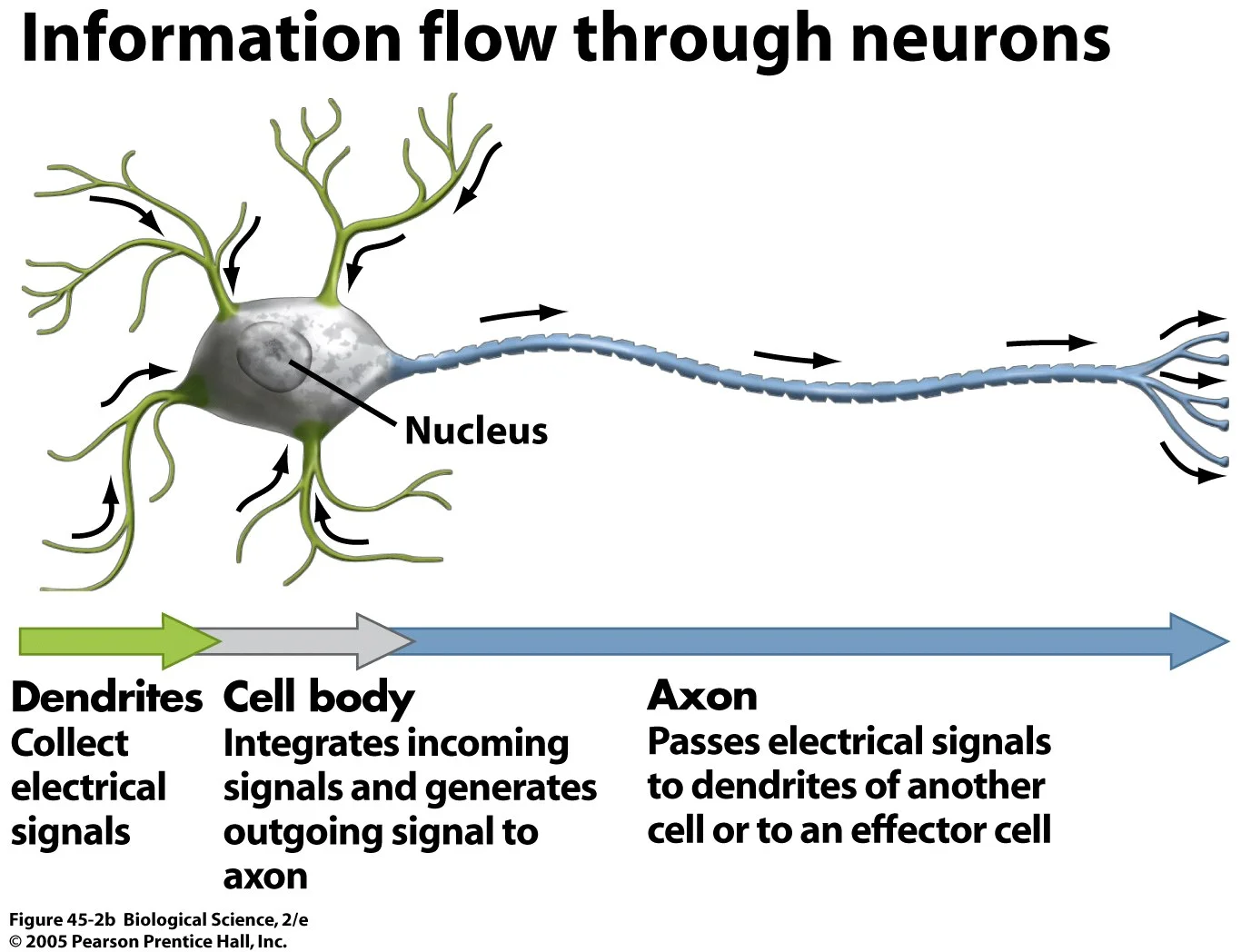
Here’s some really interesting information that may change our traditional views about information flow in the nervous system. Neuroscience students learn early on about the mechanics of nerve impulses. It’s important because it’s how neurons, the cells of the nervous system, convey information to each other.
Feel free to start reading after the figure to get the good stuff, but this simple summary of action potentials might be useful to understand it. A neuron consists of a cell body with dendrites and an axon extending from it. Dendrites receive information from another cell’s axon terminal. Each connection between two cells is called a synapse; the cell sending information is referred to as the pre-synaptic cell, and the cell receiving the information is called the post-synaptic cell.
The neuron’s membrane acts as a barrier between the outside and inside of the cell, maintaining a difference in electrical charge between the inside and outside of the neuron. The inside has a more negative charge compared to outside of the cell. This electrical difference is called the resting potential.
The pre-synaptic cell releases chemicals called neurotransmitters to the post-synaptic cell at the synapse. Neurotransmitters can have an excitatory or an inhibitory effect on the post-synaptic cell’s charge. If the cell receives inhibitory neurotransmitters, the inside of the cell becomes more negative (hyperpolarization). On the other hand, if the cell receives excitatory neurotransmitters, the inside of the cell moves toward a more positive charge (depolarization).
Each cell has a threshold of excitation. When the inside of the cell reaches that level after receiving enough excitatory signals, the membrane will permit the flow of positive ions into the cell, causing the electrical charge inside the cell to increase to a positive charge very rapidly. This is called an action potential.
The action potential begins at the axon hillock, which is where the cell body meets the axon. It then travels down the axon away from the cell body. In healthy mammals, the initiation of the action potential has always been observed at the axon hillock.
A recently published paper by Sheffield and colleagues asserts that in certain rodent hippocampal inhibitory neurons, some action potentials began at the distal end of the axon (the end not connected with the cell body) instead of at the axon hillock, and also that axons could communicate with each other without the signal first going through dendrites or cell bodies. While this has been observed in invertebrates, this has never been documented in mammals.
The authors stimulated these neurons in brain slices with hundreds of action potentials over tens of seconds to minutes, which resulted in persistent action potential firing in the distal axon which could last from tens of seconds to minutes. The persistent firing occurred with natural and unnatural patterns of input, and even when the inside of the cell body was maintained at a hyperpolarized level to ensure that the firing wasn’t a result of the cell body depolarizing from repeated stimulation of the distal axon.
In some pairs of neurons, stimulation to one neuron would induce persistent firing in the unstimulated neuron, which indicates communication between axons without needing the dendrites or cell bodies and without synaptic connection between axons.
Persistent firing was observed in these specific neurons of mice and rats, which shows that it occurs across species. The fact that it was only observed at temperatures of 30-37°C (86-100.4°F), closer to physiological temperatures, and that it resulted from natural patterns of stimulation may indicate that persistent firing in distal axons would occur in a whole, live organism.
What might some implications be? As mentioned earlier, persistent firing was observed in certain inhibitory neurons of the hippocampus, which is believed to be important for consolidating short term memories to long term memories. According to Sheffeld et al., it could be a mechanism to store information over short periods of time, such as for working memory. Additionally, the frequency of persistent firing closely matches beta and gamma oscillations, which are thought to play a role in cognitive processing and some psychiatric disorders. Perhaps the synchronization of persistent firing in these neurons could contribute to these oscillations. Finally, the authors speculate that the persistent firing of these inhibitory cells could be a protective mechanism against runaway excitation that occurs in seizures.
